


Our Product
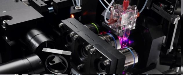
The Cytek® Amnis® ImageStream®X Mk II imaging flow cytometer combines the high throughput performance of flow cytometry with the imagery and functional insights of microscopy. This unique combination enables a broad range of applications that would be impossible using either technique alone.
The ImageStreamX Mk II system is a benchtop, multispectral, imaging flow cytometer designed for the rapid acquisition of millions of cells with up to 12 channels of cellular imagery. It collects large numbers of digital images, performs spectral compensation and provides high quality images of every cell in your sample. Combined with our advanced image analysis software where each object is quantified for hundreds of parameters using not only fluorescence intensity but the spatial location of that fluoresced as well, the ImageStream system provides an unprecedented level of cellular information.
See How Imaging in Flow Has Changed Research at Roswell Park Comprehensive Cancer Center (RPCCC)
At the RPCCC Flow and Image Cytometry facility, Drs. Paul Wallace, Hans Minderman, and staff provide users with the best tools available for increasing statistical power and throughput for applications like measuring NFkB translocation that were formerly performed by laborious microscopy. Watch to learn how Amnis protocols have helped RPCCC researchers detect rare events like circulating tumor cells.
So Many Parameters
Exceptional Sensitivity
Novel Applications
| Performance Characteristics | 40x Magnification | 60x Magnification | 20x Magnification |
|---|---|---|---|
| Numeric Aperture | 0.75 | 0.9 | 0.5 |
| Pixel Size | 0.5 x 0.5 µm | 0.3 x 0.3 µm | 1.0 x 1.0 µm |
| Field of View | 60 x 128 µm | 40 x 170 µm | 120 x 256 µm |
| Imaging Rate | 2,000 Obj/Sec | 1,200 Obj/Sec | 5,000 Obj/Sec |
Sample Characteristics
- Volume: 20-200 ul
- Utilization Efficiency: up to 95%
Physical Characteristics
- 35" W x 26" H x 25" D (889 mm x 660 mm, 635 mm)
- 400 lbs. (182 kg)
Automated Instrument Operations
- Start up and shut down
- Sample load, acquisition, compensation, batch analysis
- Laser alignment, focus, calibration, and test
Illumination
- Excitation: Standard 488 nm; Optional High Power 488 nm, 375 nm, 405 nm, 561 nm, 592 nm, and 642 nm
- Side Scatter: 785 nm standard
- Brightfield: Customizable in any channel
Operational Requirements
- 450 W, 100-240 VAC, 50/60 Hz
- No external air or water required
Cytek® Amnis® ImageStream®X Mk II Kits
| Product | Category | Part Number |
|---|---|---|
| Amnis® NFkB Translocation Kit | Cell Pathway | ACS10000 |
| Amnis® Protein Aggregation & Silicone Oil Detection Kit | Drug Discovery | ACS10001 |
| Amnis® SpeedBead® Kit for ImageStream®X System, ISX400041 | Calibration | CN-0440-01 |
Options
The ImageStream® system offers field upgradeable options for a configuration customized to your experiments.- 6 or 12 image channels
- MultiMag 20x 40x 60x objectives
- High Gain Mode
- Extended Depth of Field (EDF)
- Autosampler 96-well plate
-
Up to 6 lasers:
- 375 nm, 405 nm, 561 nm, 592 nm, 642 nm
- High powered: 400 mw, 488 nm


Software Options
IDEAS® Image Analysis Software: IDEAS software is powerful and easy to use, allowing users to create publication ready figures of their ImageStream® and FlowSight® data. The software combines familiar tools from cytometry and microscopy software including dot plots, histograms, stats tables, image gallery display optimization and more.
- Wizards for easy-to-follow standard workflows such as Apoptosis, Cell Cycle-Mitosis, Co-localization, Internalization, Nuclear Localization, Shape Change, Spot Counting and the Feature Finder wizard that allows researchers to find the best feature for their unique experiment
- Compensation wizard that applies spectral crosstalk correction for each pixel in the file based on single color controls
- 86 features per channel measuring both intensity and morphology information
- 22 function masks that identify sub-cellular components and refine the region of interest
- Customizable statistics tables
- Templates that facilitate repeat experiments and standardize analysis for all samples
- Batch processing for automated compensation and processing of each sample
Machine Learning (ML) Module: ML is a plug in for the IDEAS software that enables generation of a novel feature specifically tailored to identify the desired cell morphology. It greatly simplifies analysis by allowing researchers to hand tag truth populations and use those images to automatically create a unique classifier that will find like images.
Compliance-Enabled Software Solutions: INSPIRE™ data acquisition software and IDEAS® data analysis software are available in 21 CFR part 11 enabled versions that allow users to meet requirements for regulated settings.
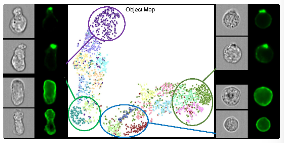
Amnis® AI Image Analysis Software: Amnis AI software represents the next generation of image analysis software. This stand-alone software package leverages computer aided hand tagging, clustering with object map plots, and generation of a novel experimental model using deep learning with convolutional neural networks (CNN) and Random Forest algorithms to classify any cell morphology. The software allows scientists to visually categorize multiple cell classes and train the computer to identify those populations in unknown samples producing easy to understand results including a confusion matrix table, probability index, precision, recall and FI statistics.
For Research Use Only. Not for use in diagnostic or therapeutic procedures.